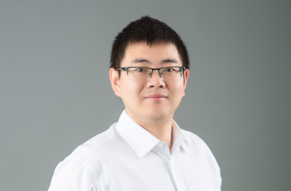
Li Lei started his master's degree in 2012 and got his Ph.D.
at the California Institute of Technology in 2019. During this period, Li Lei dedicated
himself to biomedical imaging and achieved
many accomplishments. So far, he has published 9 high-impact papers in top
journals (6 first-authored, 7 have been published, and 2 have been accepted for
publication), 4 in high-level journals (3 first-authored), 19 in optical
journals (5 first-authored), and 9 in international conferences (5 first-authored).
He applied for 4 US patents and was also invited to deliver keynote presentations on
photoacoustic tomography at the National Institute of Standards and Technology and
the National Institutes of Health.
Now he continues his postdoctoral research in Prof. Wang
Lihong's, a member of the National Academy of Engineering, laboratory at the California Institute of Technology.
Li Lei has been working on cutting-edge imaging fields. He has
developed the most advanced photoacoustic computed tomography system, named single-impulse
panoramic photoacoustic computed tomography. He demonstrated the structural and
functional imaging of the whole brain in rats and revealed the resting-state
functional connectivity of the whole brain. This is beyond the reach of
existing optical imaging techniques and provides a powerful tool for further
study of neural activity in the deep brain and the whole brain.
Li Lei combined the photoswitchable phytochrome probes to
photoacoustic tomography. He used near-infrared light to switch the photosensitive
protein on and off. By taking images at both the on and off states, the
photosensitive proteins will be selectively imaged at a very high contrast
through differential imaging of the two states. It achieved ultrahigh detection
sensitivity, high contrast, and multiscale imaging for the first time.
Li Lei also proposed a new decay characteristic analysis method,
which can realize accurate quantitative multi-contrast imaging in the deep
tissue of the living body. At the same time, the combination of the advanced
photoswitchable probes and photoacoustic computed tomography has, for the first
time, achieved real-time high-resolution imaging of protein-protein
interactions in deep tissue. This technical advancement paves a new avenue for
cancer study and drug development.
Li Lei's third academic contribution is that he has introduced the concept of ergodicity into photoacoustic imaging and realized
snapshot widefield high-throughput imaging. This technique captures wide-field
images with single laser shots by encoding spatial information with randomized
temporal signatures through the ergodic cavity. This ergodic cavity based
photoacoustic imaging system can be easily miniaturized toward wearable
applications for tumor detection and human vital sign monitoring.


