Photo of Christine Fleming

Biotechnology & medicine

Christine Fleming

Images of the beating heart could make it easier to detect and treat heart disease.
Photograph by Beth Perkins; micrographs courtesy of © 2008 Society of Photo-Optical Instrumentation Engineers, Christine P. Fleming; Crystal M. Ripplinger; Bryan Webb; Igor R. Efimov; Andrew M. Rollins “Quantification of cardiac fiber orientation using op

Year Honored
2013

Region
Global

Christine Fleming is trying to give cardiologists a powerful new tool: high-resolution movies of the living, beating heart, available in real time during cardiac procedures. Such technology might also one day help physicians pinpoint the source of dangerous irregular heart rhythms without invasive biopsies. It could even help monitor treatment.

Her invention uses optical coherence tomography (OCT), a technique that captures three-dimensional images of biological tissue. A specialized catheter with a laser and small lens near its tip is threaded through the arteries. When the laser light reflects off the heart tissue, it is picked up and analyzed to create an image. OCT has a higher resolution than ultrasound and captures images faster than magneticresonance imaging, or MRI. But today OCT has limited cardiac application—usually to search the arteries for plaques. Fleming, an electrical engineer who joined the faculty at Columbia University this year, has designed a new type of catheter capable of imaging heart muscle.

One of the primary uses of the technology will be to locate, and monitor treatment for, irregular heart rhythms that are typically caused by disruption of the heart’s regular tissue structure. In patients with arrhythmias, which can lead to heart failure, surgeons often burn away the affected tissue with targeted radio-frequency energy. Currently they perform the procedure somewhat blind, using their sense of touch to determine when they have come in contact with the muscle wall. “Since the physician doesn’t have a view of the heart wall, sometimes the energy is not actually being delivered to the muscle,” says Fleming, who adds that the procedure can last for hours. Fleming has shown in animal tests that her catheter, which uses a novel forward-facing lens, can successfully monitor the ablation in real time. Algorithms that help distinguish untreated from treated tissue offer further guidance.

Fleming is also developing algorithms to help improve the detection of arrhythmias by precisely measuring the three-dimensional organization of heart muscle. The technique works best when the tissue has been chemically treated to make it clearer, and thus easier to image. But her team at Columbia is now improving the algorithms so that the method works without this treatment. She hopes that in time the technology could supply an alternative to invasive biopsies, which are sometimes used to diagnose unexplained arrhythmias or to monitor heart health after transplants.

Fleming’s arrival at Columbia earlier this year was something of a homecoming. As a high-school student in New York City, she interned at the NASA Goddard Institute for Space Studies, which is down the street from her current lab. But in the intervening years her engineering interests have increasingly become tied to medicine; her inspiration for studying the electrical properties of the heart came when she studied electrical engineering and computer science as an undergraduate at MIT. Working with physicians is especially exciting, she says, because “you get the sense that one day your technology will be used.”

Emily Singer

inventors flemingx519

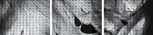
Abnormal orientation of cells in the heart wall is a clue to arrhythmias, which can be fatal. These images, created using optical coherence tomography, show the orientation of a rabbit’s heart-muscle cells. Christine Fleming’s approach to diagnosing arrhythmias could be an alternative to invasive biopsies.